She underwent prolonged periodontal treatment to manage her aggressive periodontal disease. Despite efforts, teeth #33 (22)- #43 (27) have been deemed hopeless due to extensive bone loss. The goal of…
19 year old healthy female, was referred with complaints of retrogenia. At the age of 17 she had an orthodontic treatment, for 2 years. After completion of orthodontic treatment, she…
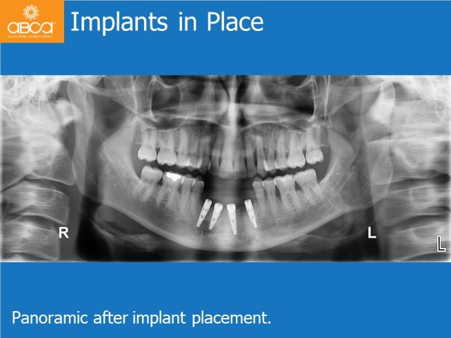
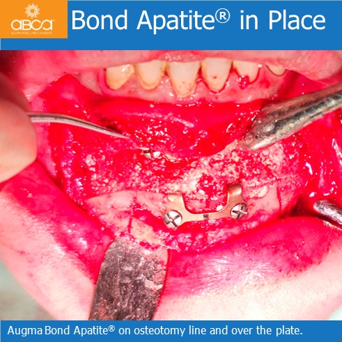
33-year-old male, otherwise healthy, heavy smoker. He was referred due to multiple cysts in the maxilla and mandible. Under general anesthesia: the patient underwent multiple extractions, cyst enucleations and bone…
The patient was a young male, after failure of crown on tooth #11 (8).
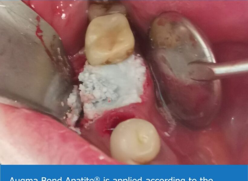
Extraction of hopeless premolars #24 (12) & #25 (13). The treatment plan included extraction and socket grafting, and the placement of two implants during a second stage. A closed sinus…
e patient was an 18 year old presenting with congenital malformation of #12 (7) & #22 (10). The patient came after opening spaces with orthodontic treatment.
Intra oral images showing failed bridge in an otherwise healthy 60 year old male. Referred to the surgeon due to recurrent inflammation and discomfort in the upper right maxilla. Periimplantitis…
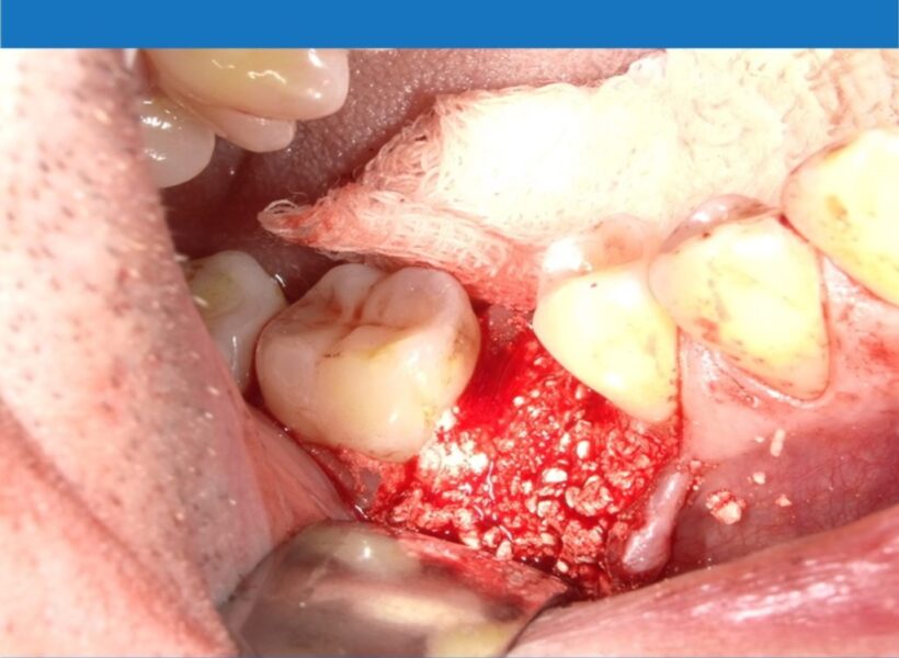
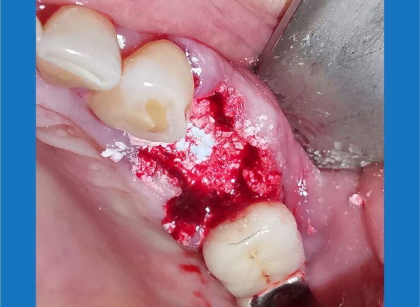
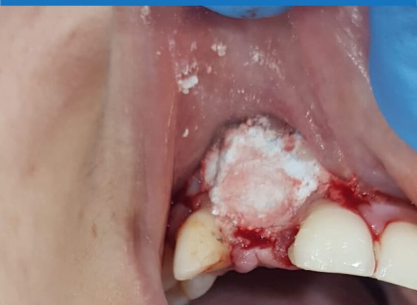
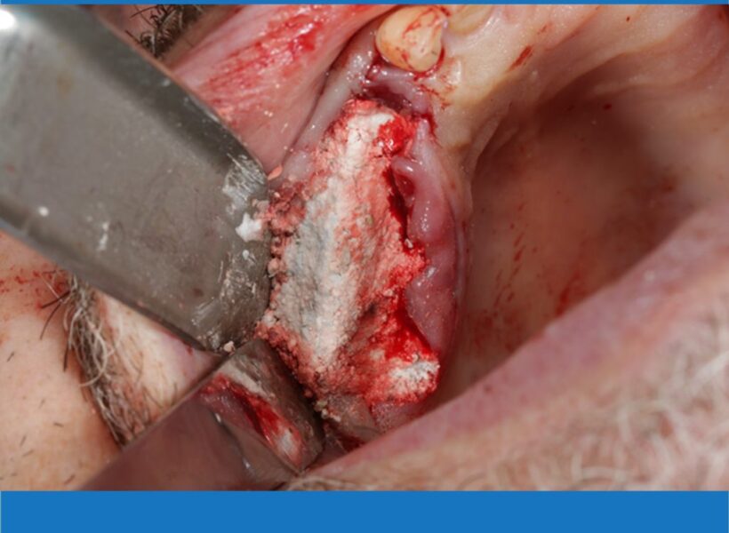
Tooth #45 (29) & #46 (30) were extracted. Tooth #44 (28) is going through an endodontic retreatment procedure. At the time of extraction, enucleation of a cystic lesion 1 cm…
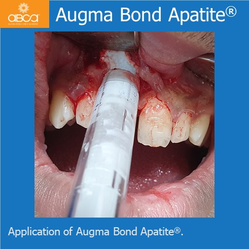
A large periapical lesion involving teeth #22 (10) and #23 (11). Cyst enucleation and apicoectomy were performed. Two year post-op photos show the entire defect filled with true bone.
The patient was referred for an implant after the extraction of tooth #12 (7). According to the CBCT, their was insufficient bone volume due to a bone defect and a…