This case demonstrates the clinical and biological versatility of biphasic calcium sulphate. All digital. The last canine remaining was used for vertical dimension of occlusion. There was no infection and…
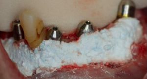
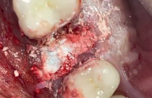
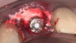
The lower left canine is both infected and fractured. Grafting was with autogenous bone and Augma Bond Apatite®. Due to uneven socket wall levels for the canine, and implant was…
and after graft placement with Augma Bond Apatite® a 6x11.5mm implant was inserted at 50n-cm final torque.
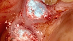
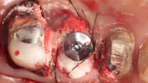
There were long standing carries in the upper left canine, so it was decided not to proceed with a socket shield. The decay extended subgingival and drilling was done at…
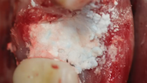
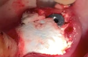
Extraction using a magnetic mallet to avoid the distal implant with flap elevation. Prep at the palatal socket relying on the interradicular bone. Augma Bond Apatite® was placed first and…
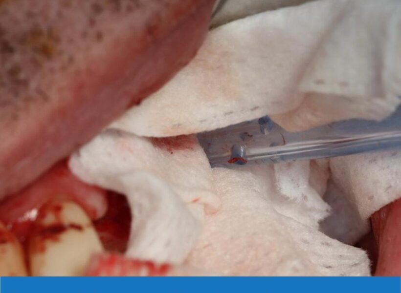
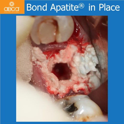
This is a sinus lift with two windows due to a partial septum. Bond Apatite® was used in the sinus lift, and was placed directly into the chamber.
The patient is 67 years old and was referred for the treatment of a fractured tooth with radiolucency. In this case there was no buccal plate and no incisions were…
The patient presented with a failed bridge, and has a history of IV bisphosphonate and extractions were completed without flaps. Socket grafting and immediate implants were placed, and the socket…
In this case there was a vertical fracture and a fistula. Extraction and immediate implant with 3D Bond.
This is a case of socket shield for the right central incisor. The left central incisors was placed 8 years prior with full extraction. One can tell the difference with…