The patient reported inflammation, swelling and pain on occasion consistent with Vincent’s symptom in the area of teeth #36 (19), #20 (35), and #34 (21). The surgery was performed under…
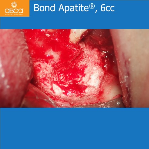
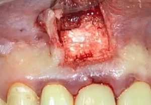
37 year old male patient presented with an apical resection with a retro filling in tooth #11 (8). The cystic cavity is filled with Augma Bond Apatite.

CASE 1 – BOND APATITE® HISTOLOGY 3 MONTHS POST OP The Biologic Effects of Bond Apatite® in New Bone Formation in Osseous Defects Bond Apatite® is a composite bone graft…

Calcium sulfate More than 100 years of documented clinical success in bone augmentation Biphasic Calcium Sulfate is composed of two phases of the well known Calcium Sulfate. Calcium sulfate (CS) features…

Due to the replacement of the cement into the patients’ own bone, the Radiographic appearance will vary during the healing period. During graft placement – Radiopaque 2-3 weeks post-op – Radiolucent 12 weeks post-op…
Sinus Lift – Intra Crestal Approach
Sinus Lift – Lateral Window Approach Activate the syringe and wait 1 minute before application. Eject the cement into the sinus cavity through the sinus lateral window until 2/3 of the sinus is filled (During…
Raise a flap The flap should be minimally reflected in order to expose the entire grafted site (Only one vertical cut should be performed no more than 2-3 mm into…

Before Flap reflection perform mesial oblique vertical incision (up to 2 mm into the mobile mucosa). Raise full thickness flap, minimally as needed to expose the entire defect (Do not perform any manipulation…
Eject the cement into the socket. Press firmly over the cement for 3 seconds using dry sterile unfolded gauze and finger pressure followed by another 3-second press with a peritoneal elevator.
