Introduction to Augma Lift™
Scenario A - < 4 mm
Scenario B - ≥ 4 mm
What to Expect
Additional Information
Augma Lift™ What to Expect – Radiographic Appearance & Soft Tissue Healing
Augma Lift™ What-to-Expect
Augma Lift™ is a minimally-invasive technique for performing intra crestal sinus lifts from 1mm height.
- Working with Bond Apatite® results in a complete bone regeneration.
- The healing process differs from many other bone substitutes, and there might be major differences in the radiographic appearance between traditional grafting and Augma bone cement grafting.
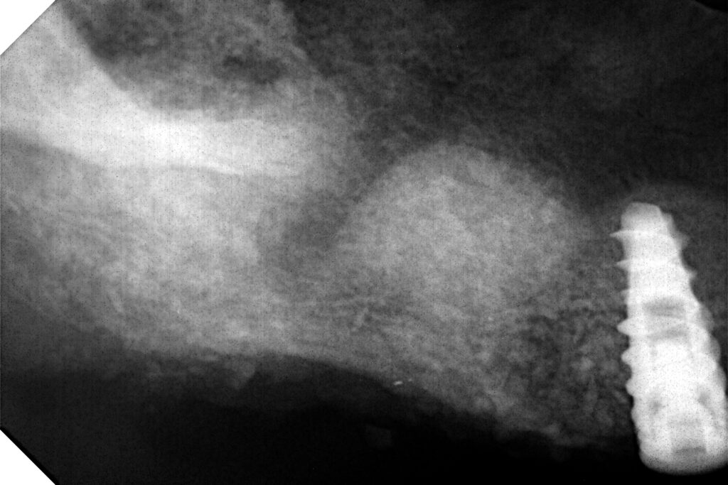
- Immediately after graft placement, the radiographic appearance is radiopaque.
- Two weeks after placement the radiographic appearance is radiolucent.
- Five months post-op it will appear radiopaque again.
- It is important to note that the radiopacity reflects the appearance of young, vital bone.
- Take into consideration that there will be some resorption and height reduction as part of the healing process.
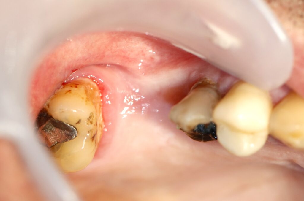
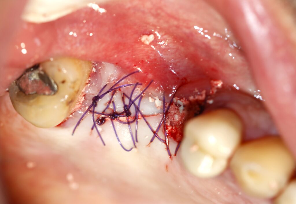
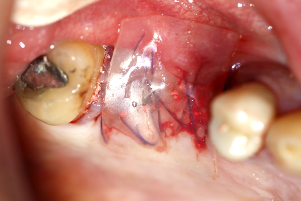
- Please view the following phases emphasizing the healing process, including soft and hard tissue healing and radiographic appearance.
Augma Lift™ | Radiographic Appearance
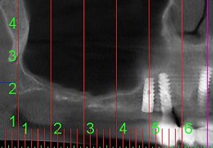
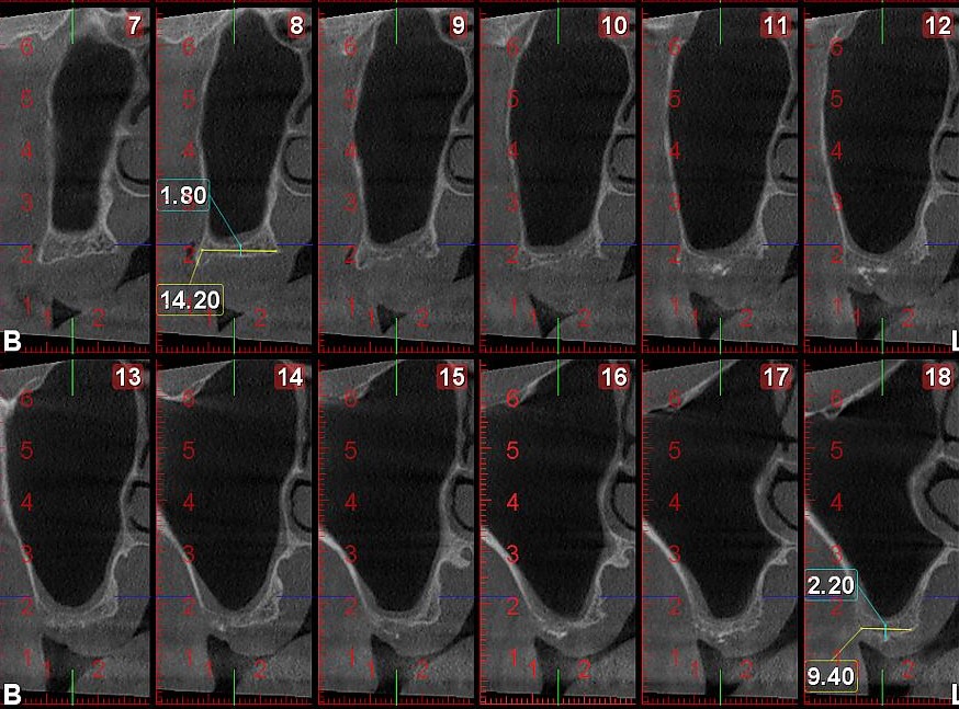
Pre-Op
Case 1 - Radiography
Immediate Post-Op
Case 1 - Radiography
4 Months Post-Op
(Prior to Implant Placement)
Case 1 - CBCT
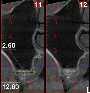
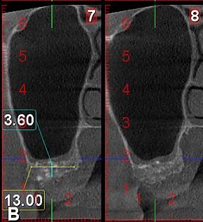
4 Months Post-Op
(Implant in Place)
Case 1 - Radiography
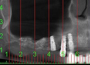
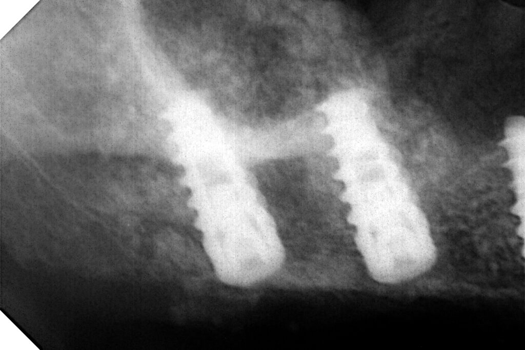
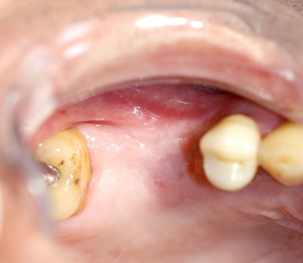
Augma Lift™ | Soft Tissue Healing
Initial Aspect
Case 2 - Soft Tissue Healing
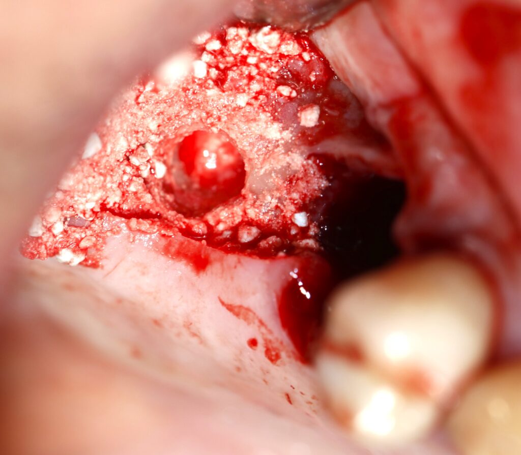
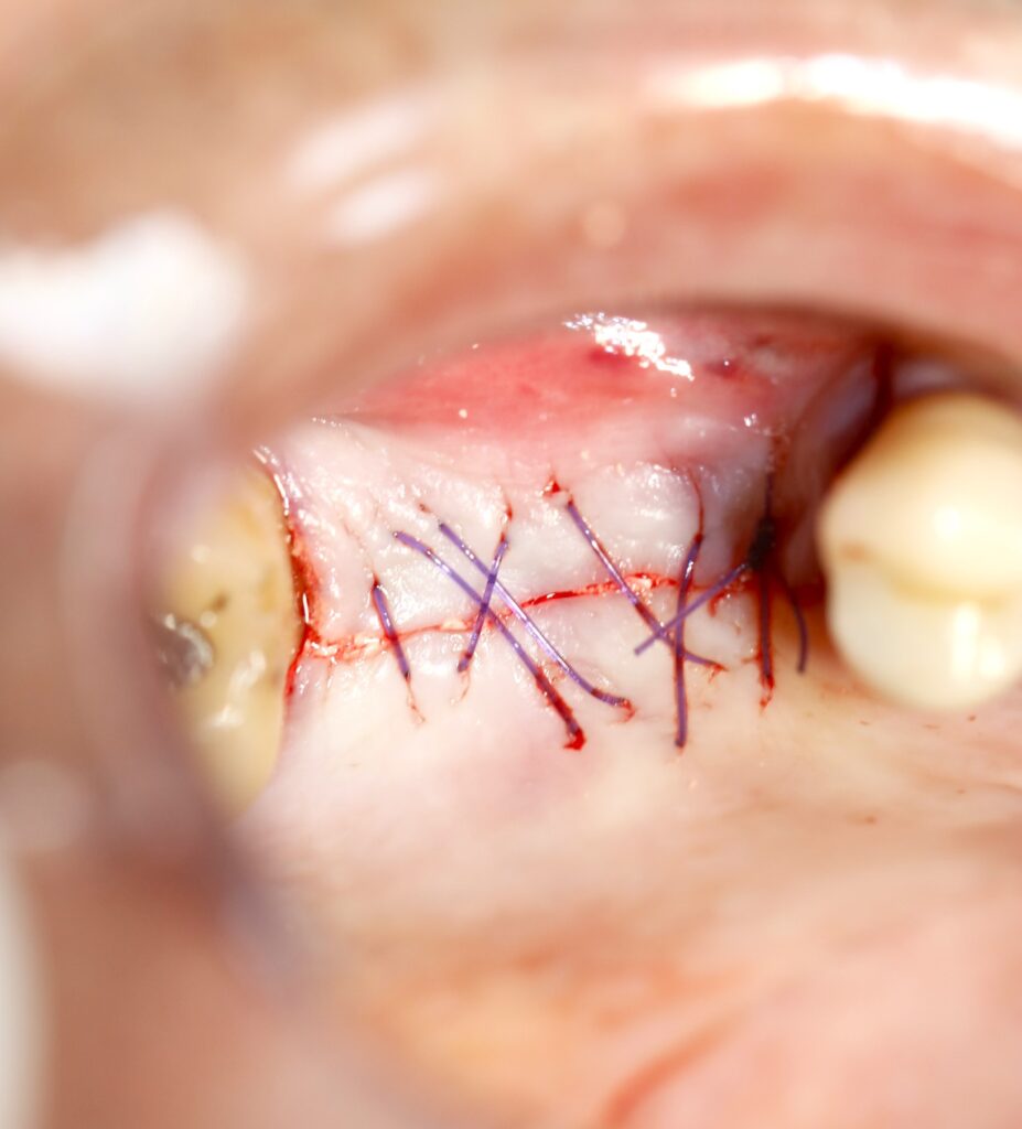
Sutures
Case 2 - Soft Tissue Healing
(Augma Shield™ was used on top of sutures as an additional layer of protection)
4 Months Post-Op
Reentry
Case 2 - Soft Tissue Healing
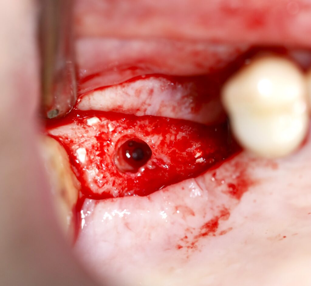
4 Months Post-Op
Implants & Sutures
Case 2 - Soft Tissue Healing
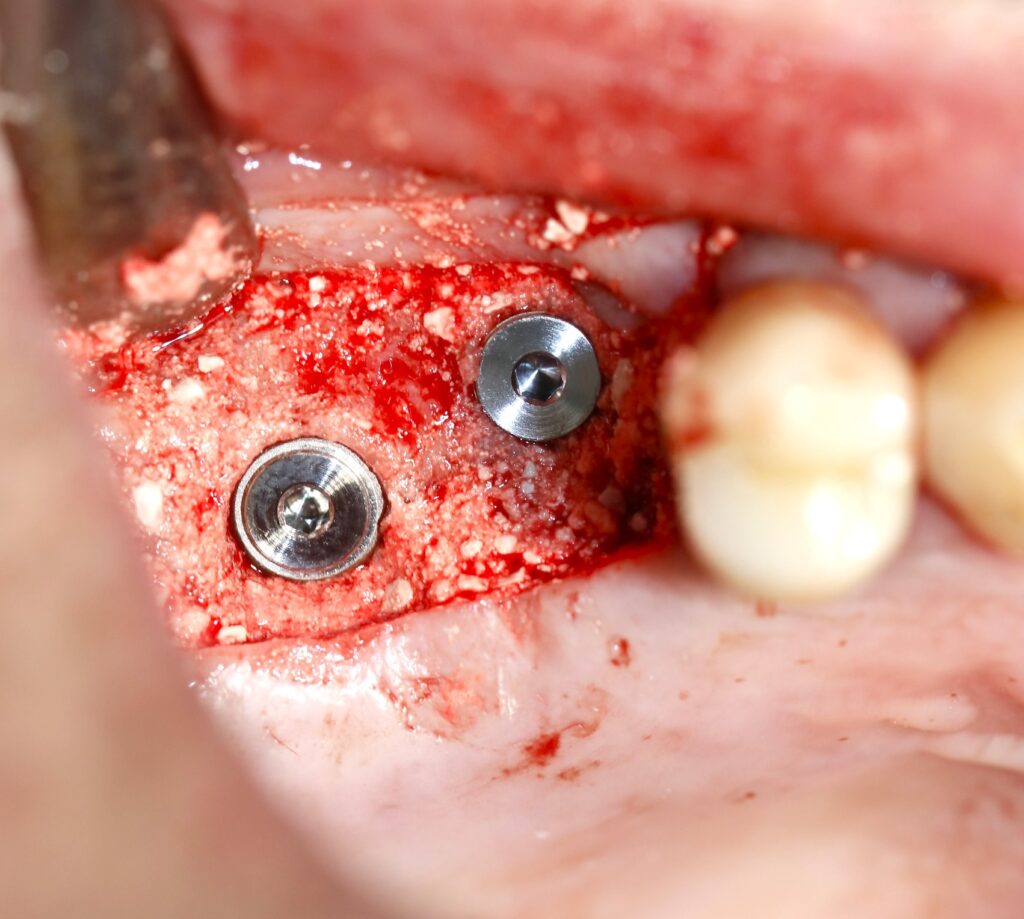
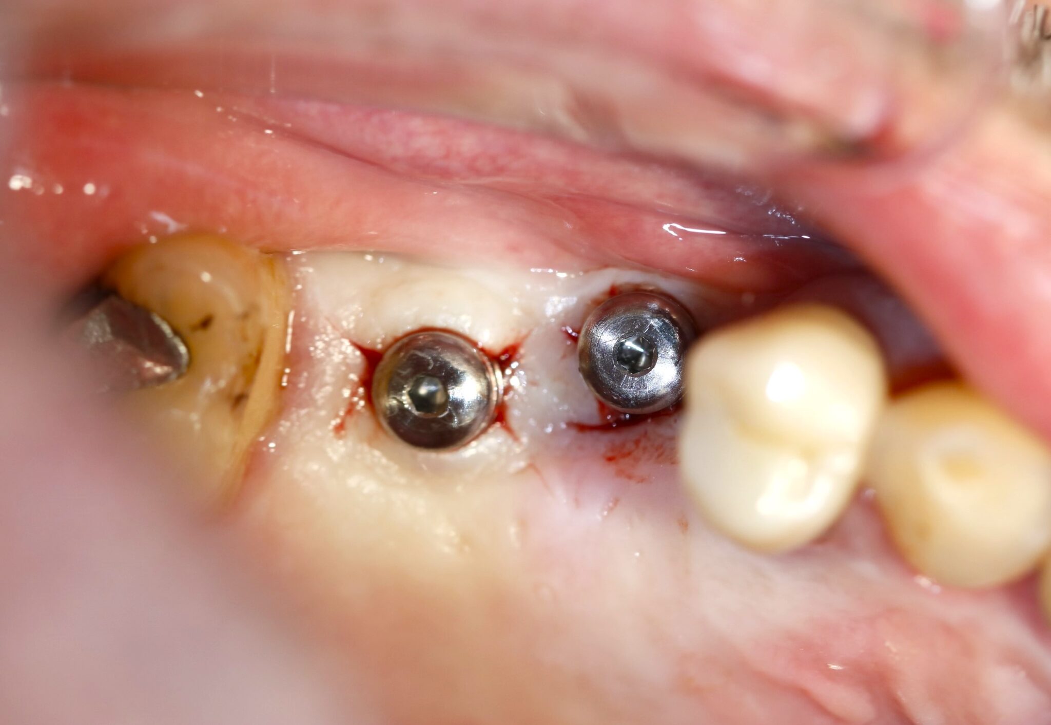
4 Months Post
Implant Placement
Case 2 - Soft Tissue Healing
