Multiple extractions, cyst removal and the reconstruction of maxillary bone defects. Rehabilitation with a temporary removable prosthesis, and 6 months post-op the final skeletal prosthesis is placed.
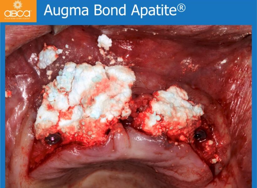
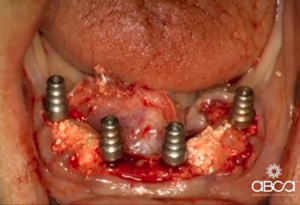
Implant placement in maxillary. The remaining teeth of the mandible are extracted, followed by ridge preservation and immediate implant placement.
The treatment will be done in stages. First, the failing central incisors will be extracted. The narrow ridge in the area of the lateral augmentation will be prepared with decortication…
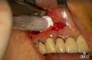
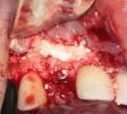
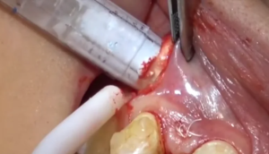
This video shows cyst removal and apicoectomy with MTA. The defect was filled with Bond Apatite and the sutures are covered with a wound dressing.
Congenitally missing maxillary lateral incisors, and deficient buccal width of alveolar bone in edentulous areas. Simultaneous graft and implant placement.
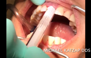
In this video see the extraction of the upper, left incisor with immediate implant placement and grafting with Bond Apatite.
In this video see the implant placement in the area of the maxillary incisors, and lateral augmentation using the tunneling technique.
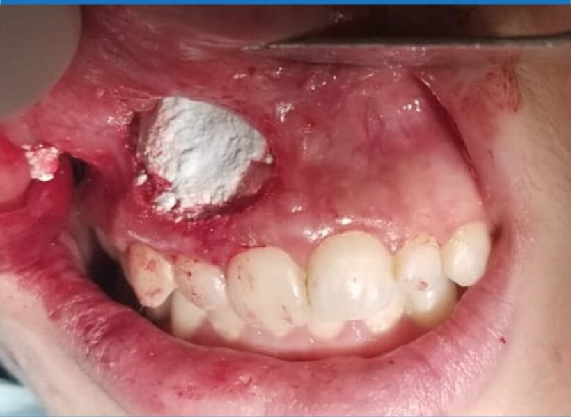
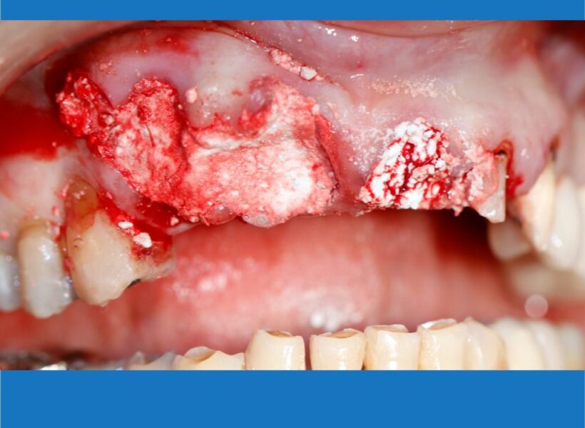
A large cyst is removed from the aesthetic zone, and the gap is filled with Bond Apatite.
The patient presented with difficulties in chewing and problems with self esteem. She no longer wanted to use a removable prosthesis and had severe maxillar atrophy.
Open to access this content