Symptomatic implant infection #14 (5), a lack of teeth for chewing, tooth mobility and a marked lack of soft/hard tissue in the edentulous area.

Open to access this content
treatment of #37 (18) and extraction of #36 (19) due to deep periodontal pocket with active secretion and furcation involvement. The tooth was extracted atraumatically, and an implant was placed…
Dr. Guy Levi

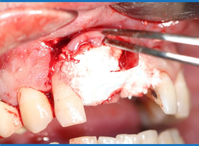
The patient presents a total failure of an old bridge between teeth #21 (9) and #23 (11) with high horizontal/vertical mobility and marked absorption of the buccal bone plate in…
Discomfort and tooth mobility of the left upper lateral incisor. A fistula was detached buccally near the apex zone of the tooth. Radiographic imagery revealed a large radiolucency in connection…
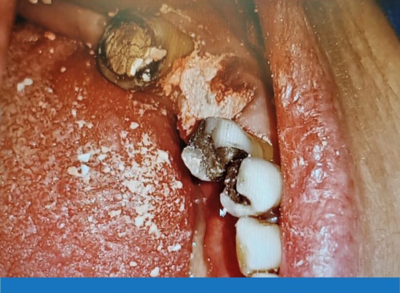
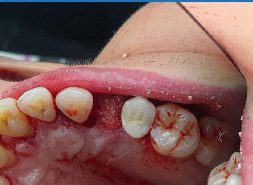
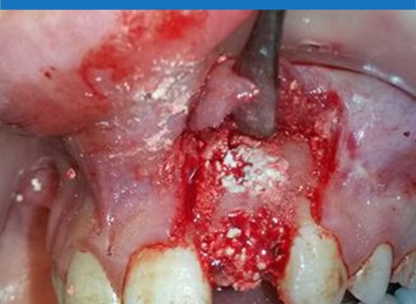
Tooth #45 (29) & #46 (30) were extracted. Tooth #44 (28) is going through an endodontic retreatment procedure. At the time of extraction, enucleation of a cystic lesion 1 cm…
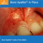
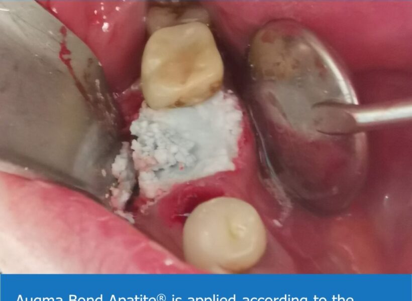
The patient presented with a crown fracture on the tooth #24 (12). The tooth was extracted and socket grafting was completed with Bond Apatite. A temporary crown was used during…
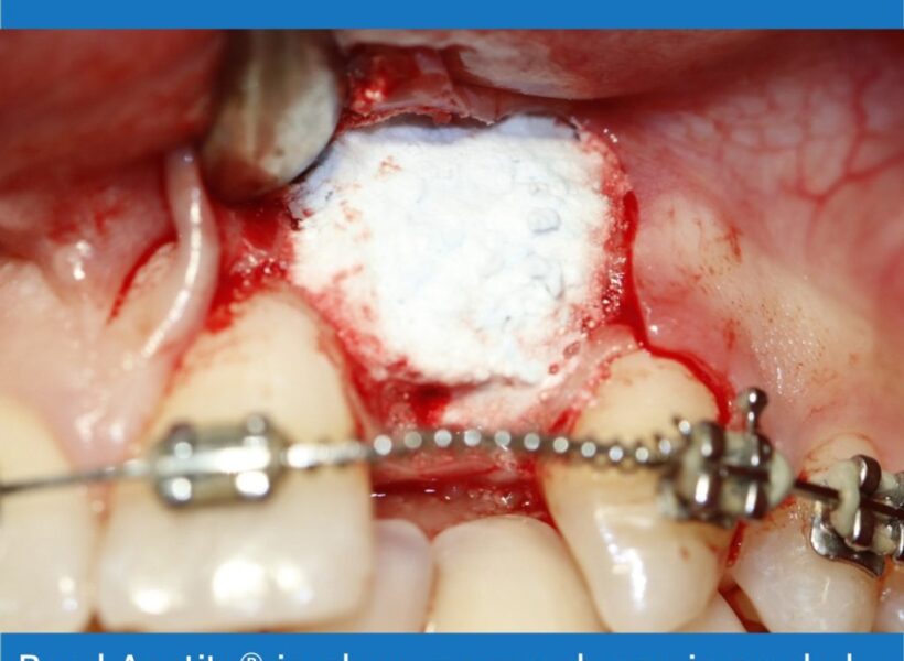
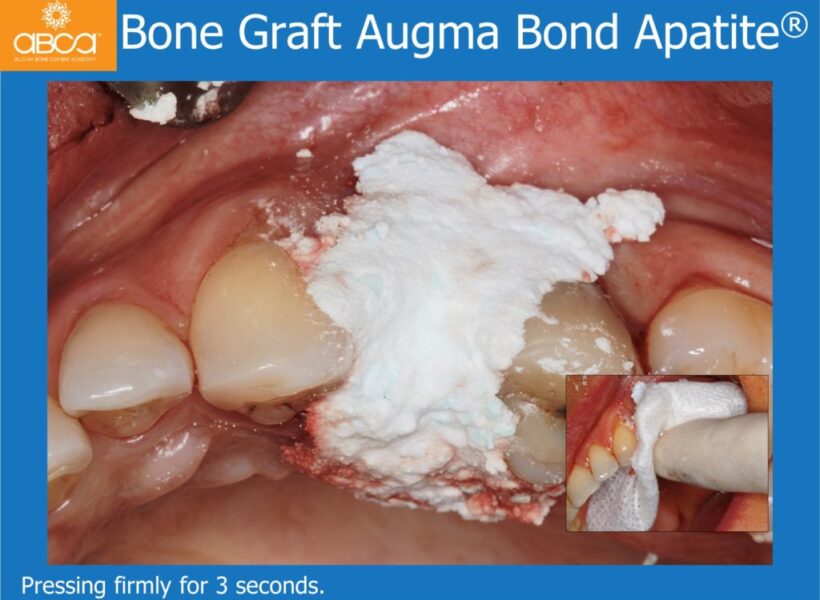
A 20 year old was referred to us to perform an implant in area #22 (10) due to agenesis (after orthodontic preparation.) The doctor wants to perform a screwed crown.…
30 year old patient presents with pain from an infection in tooth #11 (8) and the decay of tooth #21 (9) beneath the crown.