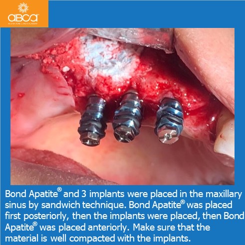
Radiographic image with absence of #14 (5) - #17 (2), and insufficient bone height for implant placement. A sinus lift should be performed. The presence of 3 mm height, from…
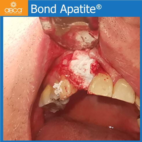
The patient was referred for an implant after the extraction of tooth #12 (7). According to the CBCT, their was insufficient bone volume due to a bone defect and a…
In this video Bond Apatite is used to fill a defect in the area of the lower, right incisor with a missing buccal plate. Four months post-op an implant is…
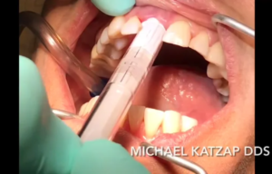
In this video see the extraction of the upper, left incisor with immediate implant placement and grafting with Bond Apatite.
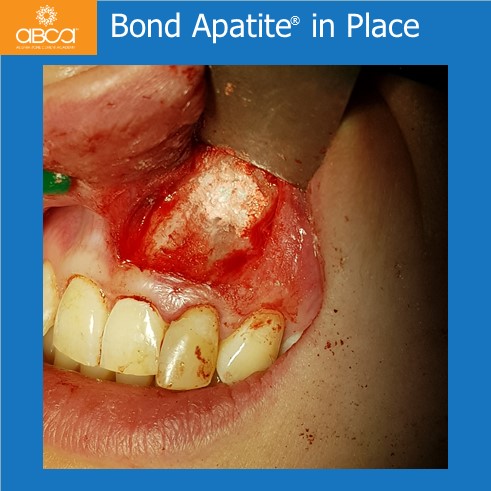
The cyst appeared 8 months after good endodontic treatment. We did a root resection of tooth #22 (10), followed by a cyst enucleation with histopathology examination. The bone defect was…
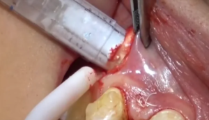
In this video see the implant placement in the area of the maxillary incisors, and lateral augmentation using the tunneling technique.
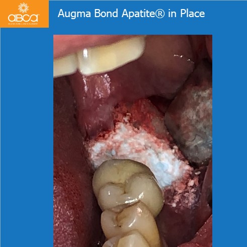
The patient returned for an unscrewing of the crown of #37 (18), which induced an occlusal trauma. This ultimately led to peri-implantitis trauma.
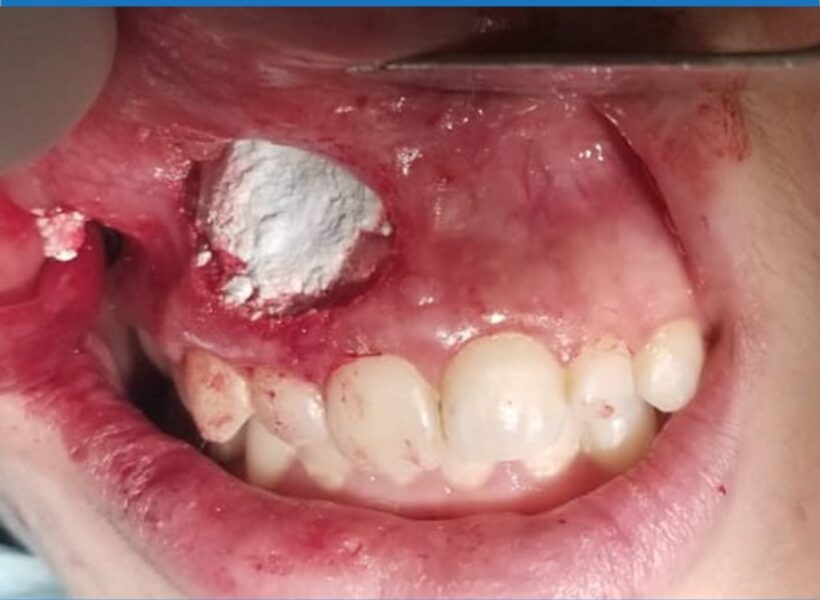
A large cyst is removed from the aesthetic zone, and the gap is filled with Bond Apatite.
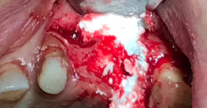
Extraction of a fractured and infected tooth. The sinus communication was repaired using 3D Bond, and Bond Apatite was then used for filling the bone defect.
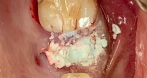
In this video the second premolar is extracted and 3D Bond is used to repair a sinus communication. Socket grafting is completed with Bond Apatite, and the graft is protected…
