Surgery David Baranes D.M.D /Amos Yahav D.M.D
- The following case represents minimally invasive, simple, and most effective surgical protocol to reconstruct a large bone deficiency in the aesthetic zone, and at the same time to preserve the papilla.
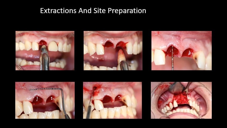
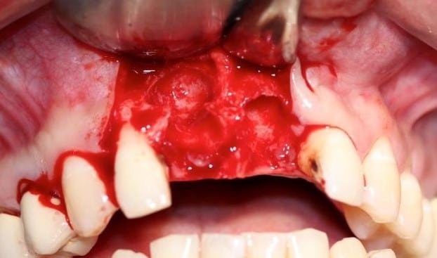
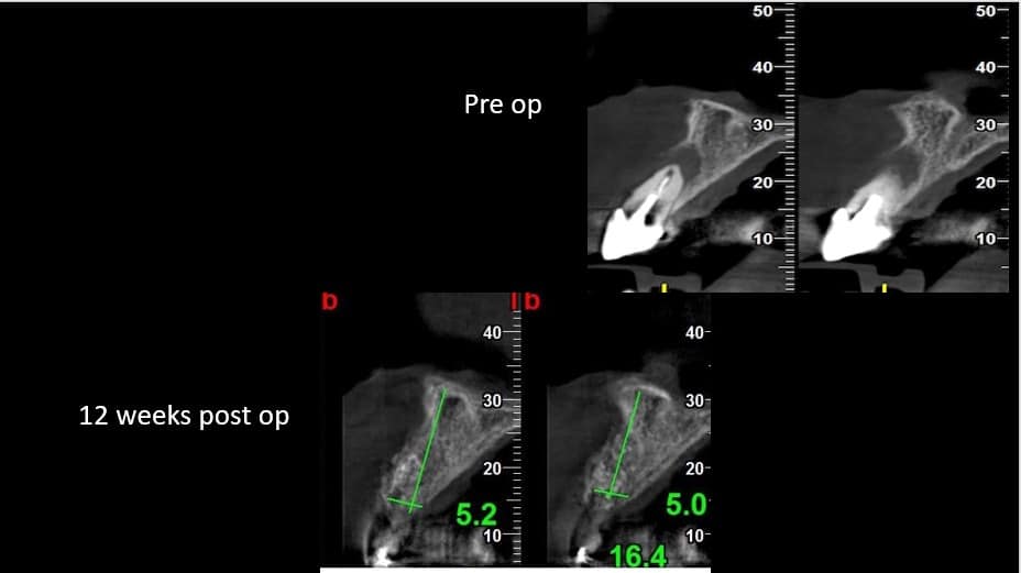
- The patient came to our office complaining of mobility and discomfort of teeth 8,9 (11,21). The clinical and radiographic evaluation confirmed the patient’s complaint. The CBCT demonstrates a large bone deficiency at the apical level with the absence of the buccal plate.
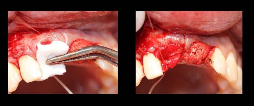
- The augmentation was performed according to minimal invasive Bond Apatite Protocols. After the extractions, one vertical incision was performed distally at a distance of one tooth from the defect site of tooth 8(11). The vertical incision does not go beyond 3 mm into the MGJ. The flap was reflected and complete debridement was done.
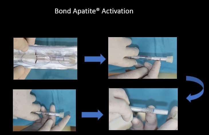
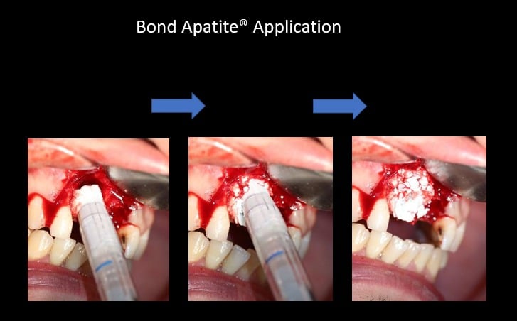
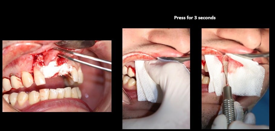
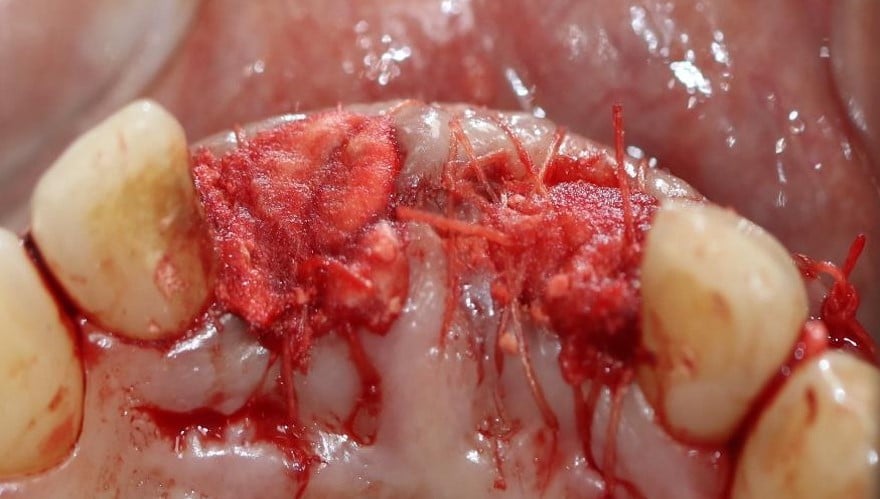
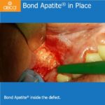
- At this stage Bond Apatite bone cement was activated and injected directly into the site, followed by compaction of the cement into place by using a sterile gauze and finger pressure for 3 seconds followed by a peritoneal elevator for additional pressure above the gauze for 2-3 seconds more on the buccal and occlusal (it is very important that the cement will be well compacted).
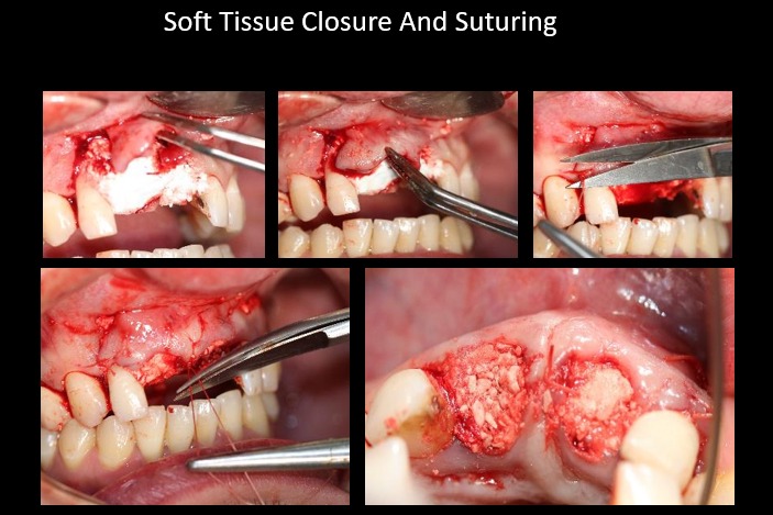
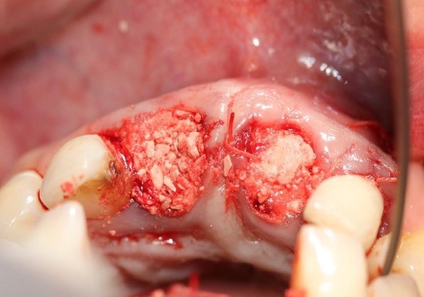
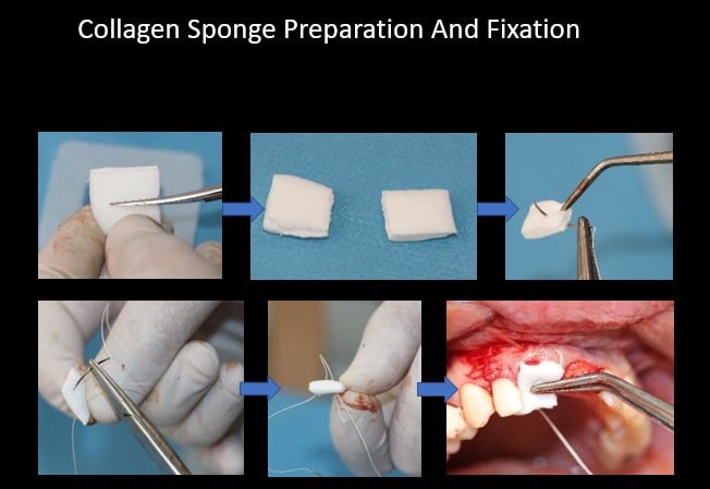
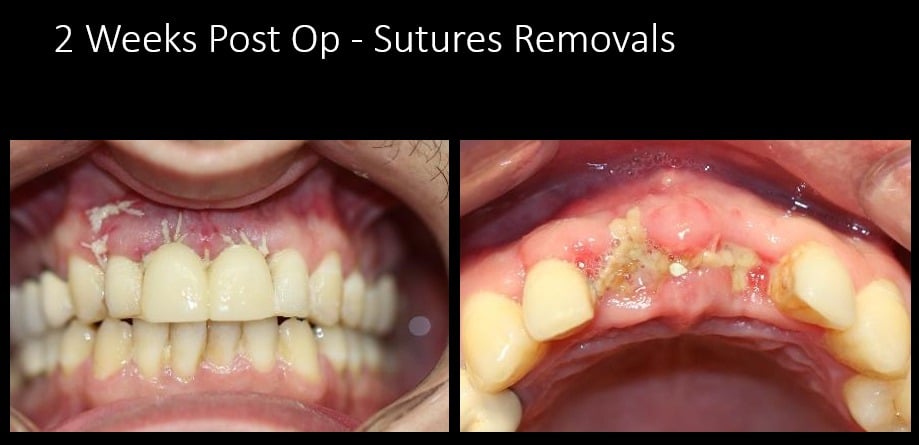
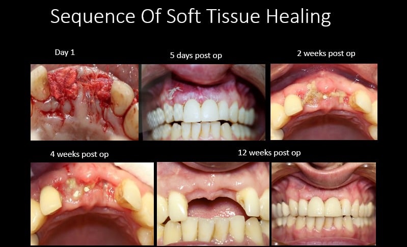
- Immediately after stabilizing the cement in place soft tissue closure took place by stretching the mesial corner of the flap and Suturing it, then the distal, then the middle portion. Due to the fact that there was one wall of a bony frame in between the two incisors, complete closure was not required and a large exposure gap can remain. However, the cement must be protected until secondary soft tissue healing takes place, therefore a simple collagen sponge was secured above.
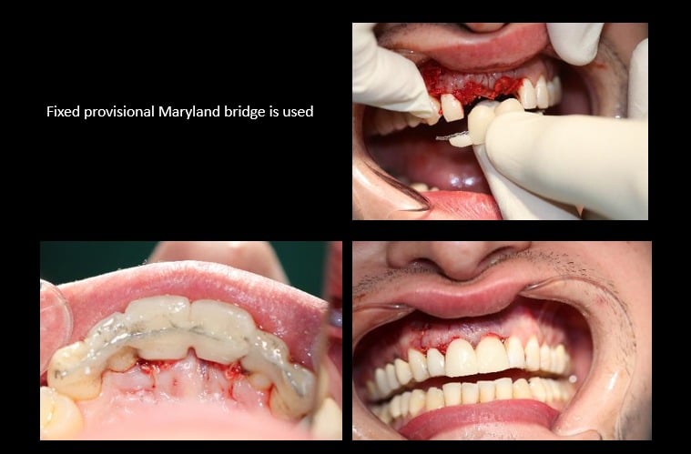
- A temporary Maryland bridge was used during the healing period.
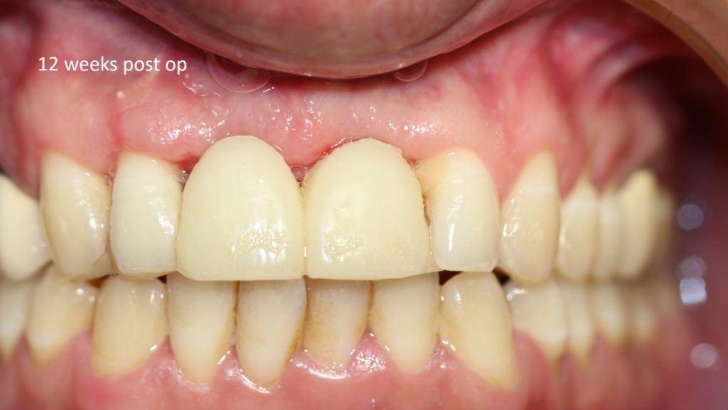
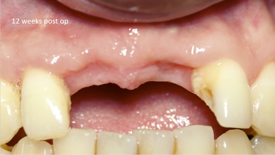
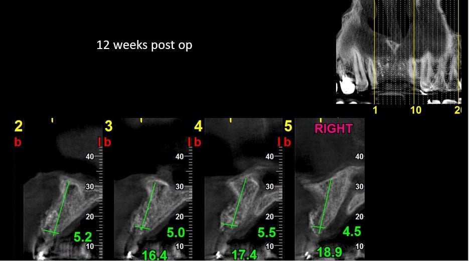
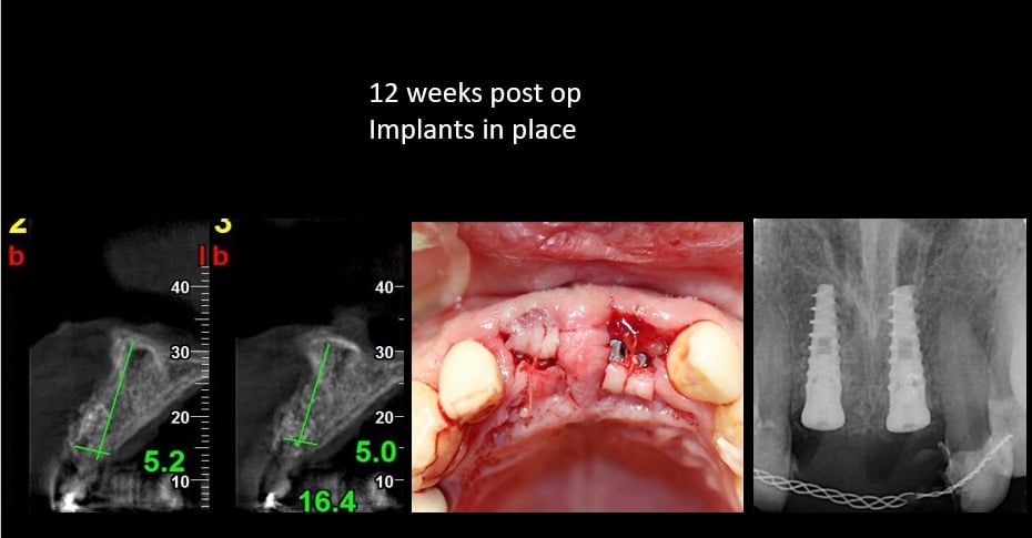
- Reentry for implant placement took place at 3 months. Healing was uneventful. Keratinized soft tissue bridged the exposed gap, the papilla was preserved in place.
- Complete bone formation can be seen clinically and radiographically.
